
A PET-CT scan is a nuclear medicine imaging technique that uses two different modalities – Positron Emission Tomography (PET)–Computed Tomography (CT) to obtain detailed information about the internal structures of the human body. For this scan, a glucose-based radiopharmaceutical called fluorodeoxyglucose (FDG), is used to localise the tissues with altered glucose metabolism. FDG accumulates in the tissue with high glucose demand, like tumours and inflammatory cells. The PET scan looks for places in the body that have a lot of glucose, this can help identify different medical conditions. A CT scan happens at the same time to show exactly where those areas are.
How does the PET-CT device look?
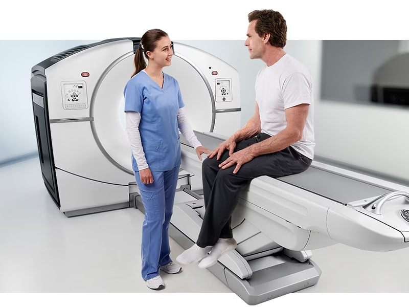
The PET-CT scanner is a cylindrical machine which comes with a tunnel at the centre. It has an additional component of a PET scanner. The gantry of the machine has an examination table that moves in and out of it. The scanner also has a technician interface with multiple monitors displaying real-time images and diagnostic information. The combination of PET and CT scan technologies allows for the simultaneous obtaining of anatomical and metabolic data.
How should you prepare?
You’ll receive all the necessary information by the nuclear medicine specialist regarding the scan. They will answer any questions or concerns you may have. You must inform your doctor and PET-CT technologist know if you could be pregnant or if you are breastfeeding. For the scan, you may be asked to:
- Drink plain, unflavoured water during this time.
- Not eat anything for 6 hours before the scan.
- Not drink tea, coffee, alcohol, squash, fizzy drinks, or flavoured water.
- Bring a list of the medications you take.
You may need to wear loose clothing that doesn’t have metal zips, buttons or studs. If your clothes have metal, you will need to change into a hospital gown.
It’s important to stay hydrated and try to drink at least three glasses of plain water before you come for the scan.
Inform the medical staff if you have diabetes.
Procedure
You will be asked to lie down on the scanner bed and keep as still as you can for the scan. Depending on the area the doctor wants to see, you may be asked to keep your arms over your head during the scan. You will then get an enhancing agent (a dye, also called contrast) through your IV.
The scanner bed will then move in toward the scanner opening for a short time to complete the CT part
of the scan. Next, the PET scan will start. The bed will once again move. The technologist can see you at all times during scanning and may give you more instructions. You should lie as quietly as you can. The scan is painless, and you should not feel anything. The entire scan will take about 30 minutes to an hour to finish, depending on the areas being imaged.
Imaging output
The images obtained through the PET scan are color-coded to depict the areas of high and low tracer concentration and high and low metabolic activity. The cancer cells appear as bright spots on the images.
The images obtained with the CT scan are highly detailed and show the body’s anatomical structures. These images are useful in identifying abnormalities in the structure of tissues and organs.
When is a PET/CT scan used?
- Cancer: to show active areas for staging.
- Brain tumours: to see if a recurrent tumour is present.
- Heart problems: to show blood flow and metabolism
- Alzheimer’s disease: to show areas of reduced activity.
- Epilepsy: to show where the seizures are coming from.
- Parkinson’s disease: to show areas of reduced function.
If you live in Delhi-NCR, you can do an online search with keywords PET CT scan price to get information regarding the best diagnostic centres in your vicinity.
Disclaimer: For personalised advice and further information, always consult your doctor or qualified healthcare professional.
